Consultant Dermatologist with 35 years experience
Author Differential Diagnosis in Dermatology
Medical and Surgical dermatology - skin rashes, moles & skin cancer
Removal of skin lesions carried out at first visit
*** IDEAL FOR NON-INSURED PATIENTS ***
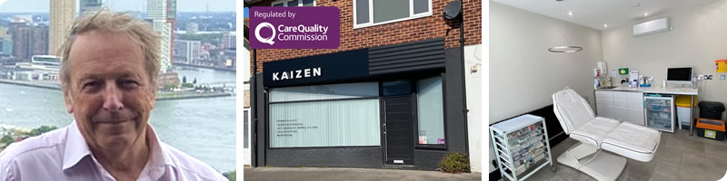
Surgery carried out in clinic room so NO extra fee charged
(unlike those charged by the Private Hospitals) |